
Ganglion Cyst Wrist: Diagnosis, Causes, and Advanced Removal
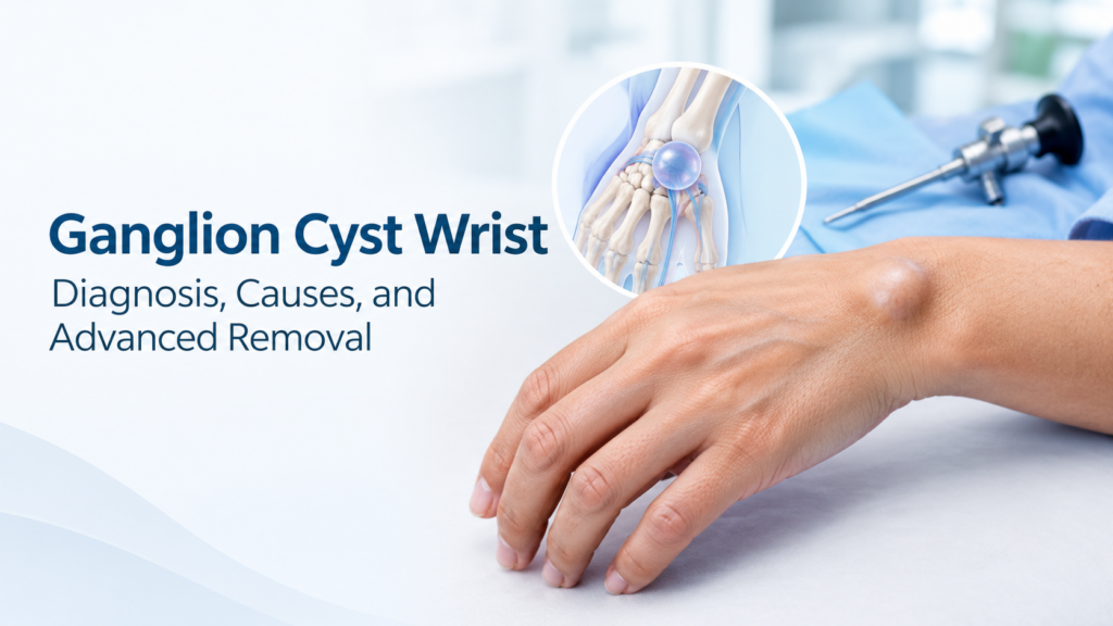
Clinically authored by Dr. Mohammad Ali, Fellowship-Trained Hand, Wrist and Upper Limb Surgeon in Dubai, UAE. A ganglion cyst wrist is the most common benign soft-tissue swelling found in the hand and wrist. Developing along joint capsules or tendon sheaths, these fluid-filled sacs often fluctuate in size and cause localized discomfort or nerve irritation. Understanding accurate anatomical origins, needle aspiration recurrence realities, and modern minimally invasive surgical options ensures you choose the most effective, lasting treatment path. Key Takeaways What Is a Ganglion Cyst Wrist and Why Does It Form? A ganglion cyst wrist represents a smooth, firm, fluid-filled swelling that emerges adjacent to a carpal joint or tendon sheath. These lumps are entirely benign and cannot turn into malignant tumors. Inside the sac is a thick, clear, gelatinous fluid high in hyaluronic acid, glucosamine, and albumin, resembling condensed synovial fluid, which means the natural lubricating liquid found inside normal joints. Anatomy of the Joint Capsule and Mucin Fluid The cyst forms when joint stress or micro-trauma causes capsular tissue to stretch or degenerate. This process creates a narrow stalk or pedicle, which means a small connecting stem, linking the joint space to the cyst. The stalk functions like a one-way check valve, allowing joint fluid into the cyst sac while preventing it from flowing back into the joint capsule. Dorsal Ganglion Cyst Wrist vs. Volar Ganglion Cyst Wrist The location of the lump dictates both the surgical approach and the anatomical structures requiring protection. Anatomical Feature Dorsal Wrist Ganglion Volar Wrist Ganglion Prevalence 60% – 70% of all wrist ganglia 18% – 20% of all wrist ganglia Typical Origin Scapholunate ligament and capsule Radiocarpal joint or STT joint capsule Visible Location Back of the wrist, prominent with flexion Palmar wrist crease near the thumb base Adjacent Structures Extensor digitorum communis tendons Radial artery, palmar cutaneous nerve Transillumination Readily transilluminates with light Transilluminates (requires vascular check) Dorsal cysts become noticeably prominent when bending the wrist downward. Volar cysts sit adjacent to the radial pulse, requiring meticulous care to safeguard blood vessels during intervention. What Causes a Ganglion Cyst Wrist to Suddenly Appear? A ganglion cyst wrist often appears without a single identifiable traumatic event. However, repetitive loading, ligament laxity, and micro-tears in the joint capsule create favorable conditions for cyst progression. Patients engaged in gymnastics, weightlifting, racquet sports, or intensive manual work show higher rates of symptomatic capsular swelling. Repetitive Wrist Strain and Capsular Weakness Repetitive extension and flexion generate micro-tears in stabilizing wrist ligaments. The body responds by producing extra mucin fluid to protect the joint. As internal joint pressure increases, this fluid herniates through weak capsular fibers to form a distinct subcutaneous pocket, which means a fluid pocket sitting just beneath the skin. Why Does My Wrist Ganglion Fluctuate in Size? Patients frequently ask why their wrist bump seems larger after busy workdays and smaller after resting. The one-way valve stalk pumps more fluid into the cyst whenever joint motion increases internal carpal pressure. During periods of wrist rest or immobilization, fluid production slows down and water content is gradually absorbed by surrounding tissues. Symptoms and Differential Diagnosis for Lumps on the Wrist While many cysts cause no pain, others produce distinct functional limitations depending on size and nerve involvement. Patients who require comprehensive clinical evaluation for chronic wrist pain treatment often present with aching during loaded wrist extension. Ganglion Cyst Wrist Pain and Nerve Tingling Discomfort from a ganglion cyst wrist stems from two primary mechanisms: How Do I Know If My Wrist Lump Is a Ganglion Cyst or a Tumor? Patients naturally worry about malignancy when discovering a sudden subcutaneous mass. True soft-tissue sarcomas in the wrist are exceptionally rare and typically feel hard, fixed to deep tissues, and completely non-translucent. Other non-cancerous lumps that can appear around the wrist include: Clinical Diagnostic Tests: Transillumination, Allen’s Test, and Ultrasound Accurate diagnosis involves three key clinical and imaging steps: 1. Transillumination Test: In a darkened room, a penlight placed against the mass reveals whether light passes clearly through mucin fluid, which means showing transillumination, the diagnostic passage of bright light through fluid-filled tissue. 2. Allen’s Vascular Patency Test: Prior to evaluating any volar cyst, the hand surgeon verifies uninterrupted arterial blood flow through both radial and ulnar arteries. 3. High-Resolution Musculoskeletal Ultrasound: Confirms the cystic, anechoic nature of the mass, maps the joint stalk, and pinpoints its exact distance from the radial artery. Non-Surgical Treatments and Home Care Realities Asymptomatic ganglion cysts do not require immediate procedural intervention. Splinting and Activity Modification When mild aching occurs during active sports or typing, conservative immobilization can calm joint irritation. Using custom orthopedic splinting to reduce joint capsular strain limits repetitive wrist extension and decreases fluid pumping across the stalk. Non-steroidal anti-inflammatory medications provide short-term pain relief by decreasing surrounding synovial inflammation. Why You Should Never Hit a Ganglion Cyst with a Heavy Book Historically termed “Bible cysts” because people struck them with heavy household books, this dangerous practice must be avoided. Smashing the cyst creates severe risks, including: Aspiration vs. Surgical Excision: Recurrence Rates and Long-Term Results When a ganglion cyst wrist causes chronic aching, weakness, or aesthetic distress, clinical intervention becomes necessary. Choosing between needle aspiration and surgical removal depends on recurrence risk tolerance, lump location, and symptom severity. Needle Aspiration Efficacy and the 50% Recurrence Reality Aspiration involves numbing the skin, inserting an 18-gauge needle, and drawing out the thick mucin jelly. Because aspiration leaves the underlying capsular stalk intact, the valve continues pumping fluid back into the cavity. Aspiration of volar wrist cysts carries added hazard due to radial artery proximity and is rarely recommended without real-time ultrasound guidance. Comparison of Wrist Ganglion Management Strategies Treatment Method Invasiveness Recurrence Rate Recovery Time Primary Advantages & Limits Watchful Waiting Non-invasive Spontaneous resolution in 30%–50% Immediate Low risk; suitable for painless lumps that do not interfere with function. Needle Aspiration Minimally invasive in-office 45% – 60% recurrence 1 – 2 days Immediate cosmetic relief; high recurrence because
Broken Hand Surgery: Fixation Options, Recovery & Care Guide

Broken Hand Surgery: Complete Guide to Procedures, Fixation, and Recovery Written by Dr. Mohammad Ali, Fellowship-Trained Hand, Wrist and Upper Limb Surgeon at Dubai Hand, providing specialized orthopedic and reconstructive extremity care in Dubai, UAE. Facing broken hand surgery can feel overwhelming, especially when finger mobility and grip strength are at risk. An accurate anatomical alignment ensures you regain full hand function without long-term stiffness. This clinical guide breaks down surgical indications, modern fixation hardware, realistic recovery timelines, and therapy protocols so you can make informed choices about your treatment. Understanding Hand Fractures: Anatomy and Common Break Patterns The human hand contains 27 bones, including 8 carpal bones in the wrist, 5 metacarpals in the palm, and 14 phalanges across the fingers and thumb. Because the hand operates via an intricate balance of extrinsic tendons, intrinsic muscles, and joint capsules, even minor bony displacement can cause severe functional limitations. Hand fractures typically fall into specific anatomical categories based on the mechanism of injury: To determine if non-operative care is viable, explore our clinical overview of custom orthopedic splinting techniques for non-displaced hand injuries. When Does a Broken Hand Require Surgery? Not every broken hand requires an operation. Many stable, non-displaced fractures heal successfully with dedicated immobilization. However, specific structural instability patterns mandate surgical intervention to prevent permanent disability, according to clinical guidelines from the American Academy of Orthopaedic Surgeons (AAOS). Clinical Indications for Surgical Fixation Orthopedic hand surgeons evaluate several radiographic and clinical parameters when deciding between conservative splinting and operative repair: Surgical vs. Non-Surgical Treatment Comparison Clinical Feature Conservative Splinting / Casting Broken Hand Surgery (Internal / Pin Fixation) Fracture Alignment Non-displaced or stable after closed reduction Unstable, displaced, rotated, or comminuted Joint Involvement Extra-articular or <1 mm step-off Intra-articular displacement >1–2 mm Immobilization Window Strict immobilization for 4 to 6 weeks Rigid fixation allows active motion in 3 to 10 days Tendon Adhesion Risk Moderate due to prolonged stiffness Significantly lower with early mobilization protocols Main Risks Secondary displacement, malunion, joint stiffness Surgical site infection, hardware prominence, nerve irritation Can a Broken Hand Heal on Its Own Without Surgery? While bone tissue will eventually generate bridging callus even when displaced, healing in an anatomically incorrect position leads to malunion. A malunited metacarpal or phalanx causes chronic pain, weak grip, altered biomechanics, and aesthetic deformity. When complex malunions occur, patients often require corrective realignment such as a corrective osteotomy procedure to restore hand alignment. Modern Fixation Techniques in Broken Hand Surgery Surgical management of hand fractures has evolved from prolonged plaster immobilization to precise, low-profile internal fixation systems that protect adjacent soft tissues. The choice of hardware depends directly on the location, configuration, and bone quality of the fracture, as outlined in clinical literature indexed by the National Center for Biotechnology Information (NCBI Bookshelf). Percutaneous Kirschner Wire (K-Wire) Pinning Percutaneous pinning involves passing smooth, surgical-grade stainless steel wires across the fracture site under fluoroscopic (live X-ray) guidance. Open Reduction and Internal Fixation (ORIF) with Mini-Fragment Plates ORIF utilizes pre-contoured, low-profile titanium plates (ranging from 1.3 mm to 2.0 mm) secured with locking or non-locking screws directly to the bone surface. Intramedullary Headless Compression Screws Cannulated headless compression screws are inserted down the central canal of the metacarpal or phalanx through micro-incisions. Because the screw heads countersink completely beneath the cartilage surface, there is no hardware friction against gliding extensor or flexor tendons. 2026 Advances: WALANT and Minimally Invasive Hand Fixation The surgical standard for hand trauma has shifted toward Wide Awake Local Anesthesia No Tourniquet (WALANT) techniques, thoroughly detailed by the American Society for Surgery of the Hand (ASSH). Using a buffered combination of lidocaine and epinephrine, surgeons achieve complete local anesthesia and field hemostasis without requiring general anesthesia, an arm tourniquet, or pre-operative fasting. WALANT allows patients to remain fully conscious and comfortably move their fingers intraoperatively. This allows the surgeon to confirm that plates and screws do not catch on tendons and verify that rotational scissoring is eliminated before closing the skin. If you have questions about perioperative steps, learn more about what to expect before hand and wrist surgery. Step-by-Step: What to Expect During the Surgical Procedure Broken hand surgery is typically performed as an outpatient day case, allowing you to return home the same day. Surgical protocols follow a clear, systematic flow to protect soft tissues and ensure rapid recovery: Detailed insights on soft tissue handling are outlined in our overview of specialized tendon repair techniques. Post-Operative Recovery Timeline and Hand Rehabilitation Bone consolidation requires biological time, but keeping joints moving is necessary to avoid post-traumatic stiffness. Structured rehabilitation principles from specialized orthopedic literature emphasize early edema management and progressive loading protocols to optimize functional recovery. Phased Hand Fracture Rehabilitation Milestones Recovery Phase Clinical Focus Allowed Activities & Exercises Protection Status Weeks 0 to 2 Edema control, wound healing, pain management Gentle active tendon gliding, uninvolved finger motion Post-op resting splint worn continuously Weeks 2 to 4 Suture removal, scar tissue desensitization Active range of motion (AROM), composite fist making Removable thermoplastic custom splint Weeks 4 to 6 Early bone bridging callus formation Gentle passive stretching, light daily activities (typing) Splint weaning for light household tasks Weeks 6 to 8 Radiographic bone union confirmed Progressive resistance exercises, putty squeezing Splint discontinued for daily routines Weeks 8 to 12+ Full functional strengthening and remodeling Heavy manual lifting, contact sports, high-impact gym Unrestricted hand usage Certified Hand Therapy Exercises Hand therapy begins early to preserve motion between healing tissue layers: Discover structured post-surgical guidance with our dedicated hand recovery exercises and tips. Potential Risks, Complications, and Hardware Removal Broken hand surgery carries high success rates, but being aware of potential risks enables early detection and treatment: To evaluate complex postoperative healing conditions, explore our specialized approaches to revision orthopedic surgery. Frequently Asked Questions About Broken Hand Surgery When does a broken hand need surgery? A broken hand requires surgery if the bone fragments are rotated (causing overlapping fingers), displaced into a joint surface by more than
Finger Pain Causes: Comprehensive Guide to Symptoms & Treatment in Dubai
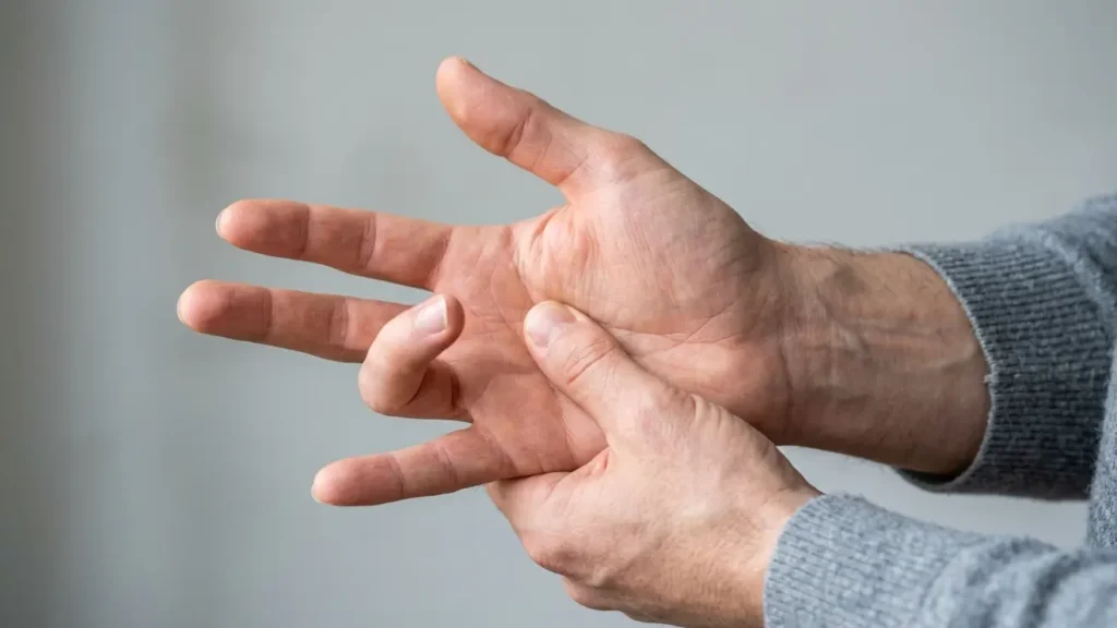
Finger Pain Causes: A Comprehensive Guide to Hand Health Authored by Dr. Mohammad Ali, a Fellowship-Trained Hand, Wrist and Upper Limb Surgeon, this guide offers trusted insights into the complex world of hand and finger health. With extensive experience serving patients across Dubai and the UAE, Dr. Mohammad Ali provides expert understanding of various conditions affecting the upper limb. Understanding finger pain causes is crucial for effective relief and treatment. Fingers are integral to daily life, and discomfort can significantly impact functionality. This comprehensive guide explores common and less frequent reasons behind finger pain, helping you identify symptoms and decide when to seek professional medical attention in Dubai. This detailed overview aims to clarify the complexities of hand discomfort. Understanding Finger Pain: A Comprehensive Overview Finger pain is a broad term encompassing discomfort anywhere from the fingertip to the base of the hand. It can manifest as a dull ache, sharp throbbing, stiffness, or burning sensation. The intricate anatomy of the finger, comprising bones, joints, ligaments, tendons, nerves, and blood vessels, means that pain can originate from numerous structures. What is finger pain and why does it occur? Finger pain occurs when these structures are damaged, inflamed, or compressed. Understanding the specific nature and location of your pain is the first step towards identifying its cause. Factors like age, activity level, medical history, and occupation can all influence the likelihood of developing certain finger pain causes. Identifying these patterns helps pinpoint the underlying issue. Common Causes of Finger Pain: Injuries and Trauma Injuries are among the most frequent reasons for finger discomfort, often resulting from accidents, sports, or falls. These can range from minor sprains to severe fractures or tendon damage. Finger Sprains and Fractures A finger sprain occurs when the ligaments supporting a finger joint are stretched or torn. Fractures, on the other hand, involve a break in one of the finger bones (phalanges). Symptoms typically include: Accurate diagnosis often requires an X-ray to differentiate between a sprain and a fracture, guiding appropriate treatment. [1] Tendon Injuries: Mallet Finger and Trigger Finger Tendons connect muscles to bones, allowing finger movement. Injuries like mallet finger happen when the extensor tendon at the fingertip is damaged, often from a direct impact, causing the tip to droop. Trigger finger, or stenosing tenosynovitis, occurs when the tendon sheath becomes inflamed and thickens, preventing the tendon from gliding smoothly. This can lead to a catching or locking sensation when bending or straightening the finger. Effective Trigger Finger Treatment is available for this condition. Cuts and Puncture Wounds Even minor cuts or puncture wounds can cause significant finger pain, especially if they affect nerves or tendons, or become infected. Deep wounds require immediate medical evaluation to assess damage and prevent complications. Inflammatory Conditions: Arthritis and Autoimmune Diseases Affecting Fingers Chronic inflammatory conditions are significant contributors to finger pain, often leading to progressive joint damage and functional impairment. Arthritis is a leading culprit. Osteoarthritis of the Finger Joints Osteoarthritis (OA) is a degenerative joint disease where the cartilage cushioning the ends of bones wears down over time. In the fingers, it commonly affects the joints closest to the fingernail (DIP joints), the middle joints (PIP joints), and the base of the thumb (CMC joint). Symptoms of finger OA often include: This condition is highly prevalent and can significantly impact dexterity and grip strength. Rheumatoid Arthritis in the Hand Rheumatoid arthritis (RA) is an autoimmune disease where the body’s immune system mistakenly attacks the lining of the joints, leading to inflammation, pain, and eventual joint destruction. It often affects multiple joints symmetrically, including those in the fingers and wrists. RA symptoms can include: Psoriatic Arthritis: Beyond the Skin Psoriatic arthritis (PsA) is a type of inflammatory arthritis that affects some people with psoriasis. It can cause finger pain, swelling, and stiffness, often leading to “sausage fingers” (dactylitis), where an entire finger swells. PsA can also affect the nails and entheses (where tendons or ligaments attach to bone), presenting a unique set of challenges for diagnosis and treatment. Gout: Sudden and Severe Joint Pain Gout is a form of inflammatory arthritis caused by a buildup of uric acid crystals in the joints. While most commonly affecting the big toe, it can also cause sudden, severe pain, swelling, redness, and tenderness in finger joints. Gout attacks can be excruciating and typically come on rapidly, often at night. [1] Nerve-Related Finger Pain: Carpal Tunnel Syndrome and Other Compressions Nerve compression syndromes are common reasons for finger discomfort, often accompanied by numbness, tingling, and weakness. These conditions occur when a nerve is squeezed or irritated. What does numbness in fingers causes indicate? Numbness in the fingers, especially when accompanied by pain, often points to nerve compression. Carpal Tunnel Syndrome is the most well-known culprit, affecting the median nerve. This condition causes pain, numbness, and tingling in the thumb, index, middle, and half of the ring finger, often worsening at night or with repetitive hand movements. Effective wrist pain treatment can often alleviate carpal tunnel symptoms. Ulnar Nerve Entrapment Less commonly, the ulnar nerve can be compressed, leading to pain, numbness, and tingling in the little finger and the other half of the ring finger. This often occurs at the elbow (cubital tunnel syndrome) but can also happen at the wrist (Guyon’s canal syndrome). Overuse and Repetitive Strain Injuries (RSI) of the Fingers Modern lifestyles and occupations often involve repetitive hand movements, making overuse and repetitive strain injuries (RSIs) significant factors in finger pain. These conditions develop gradually, often worsening over time. Tendinitis and Tenosynovitis Tendinitis is the inflammation of a tendon, while tenosynovitis refers to inflammation of the tendon sheath. Both can cause pain, tenderness, and swelling, particularly with movement. Common examples include De Quervain’s tenosynovitis, affecting the thumb side of the wrist, which can radiate into the thumb and fingers. What causes sudden finger joint pain from overuse? Sudden finger joint pain from overuse often stems from acute inflammation of tendons or joint capsules due to unaccustomed or excessive